

Table of Contents
The moment you hold the pregnancy test stick in your hand and it shows those two pink lines, there’s not only a gush of fluids that sing hallelujah, but an expecting mom also prepares herself to set foot on the upcoming journey of birthing. While the uterus swings 360-degrees, we’re sure you might be very curious as to what happens inside. What better way to know and monitor the unborn baby’s growth than through an ultrasound?
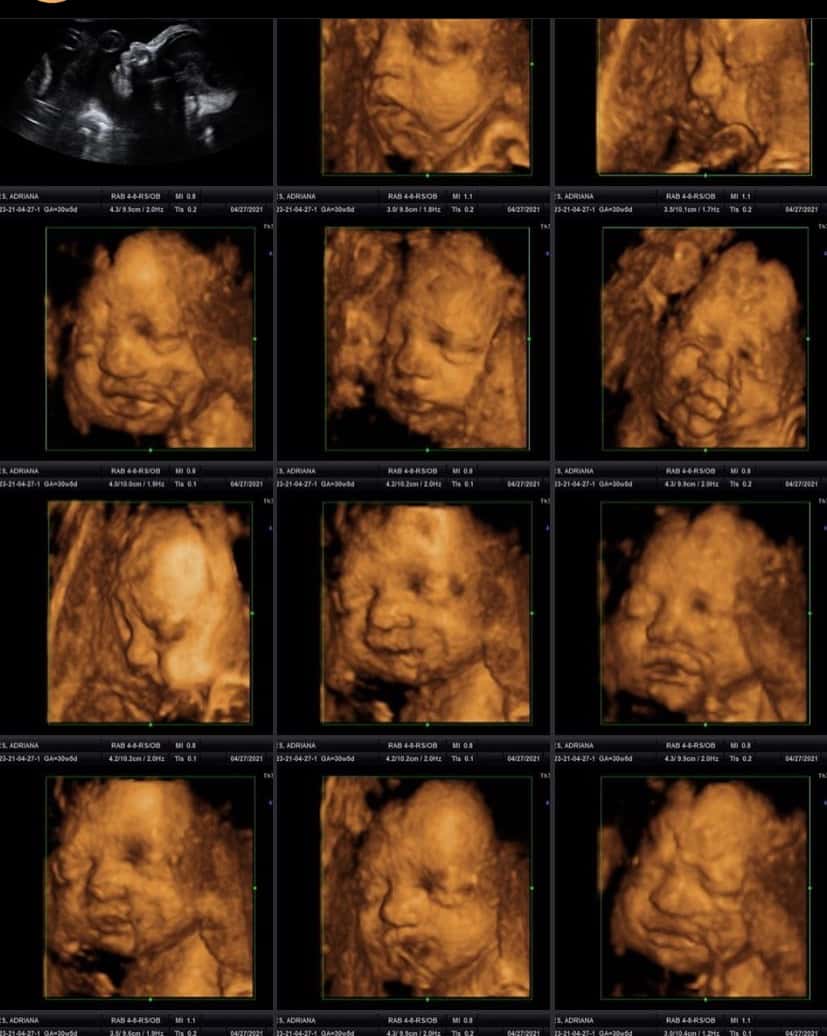
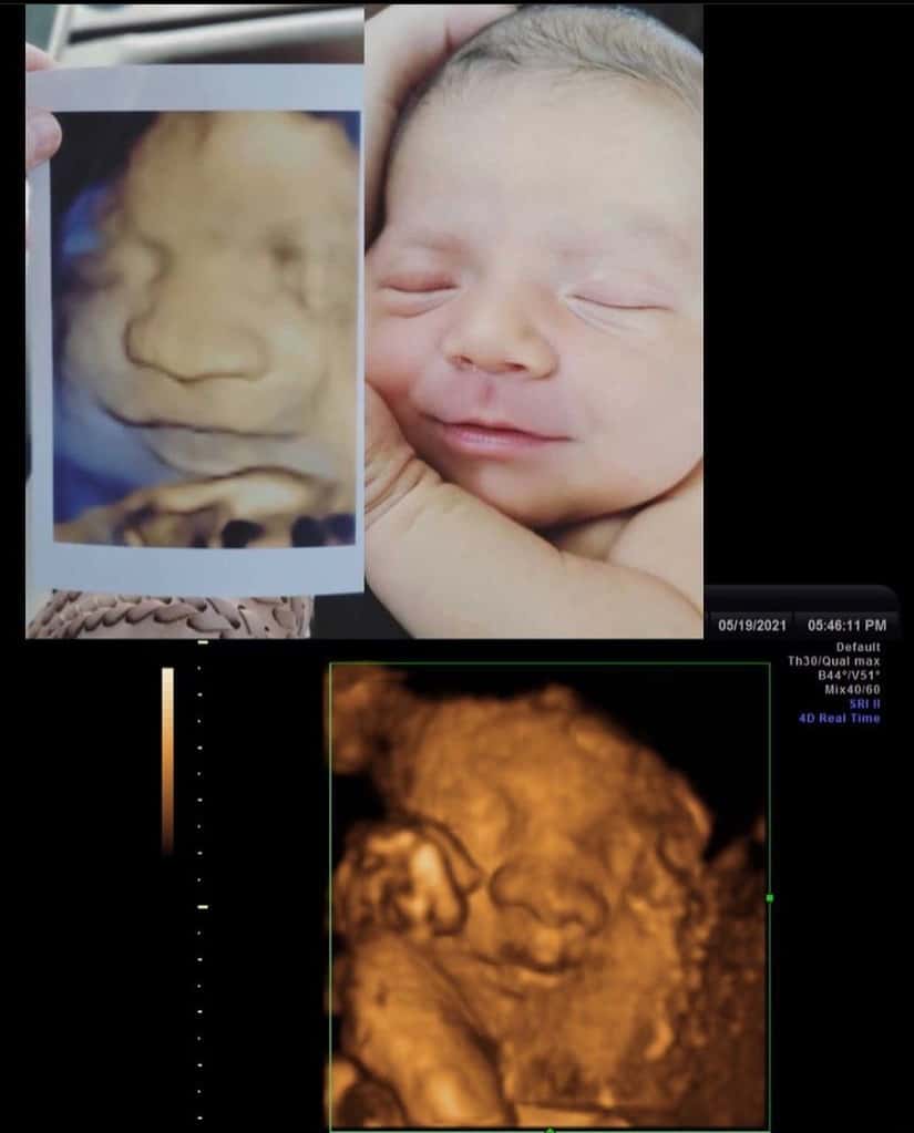
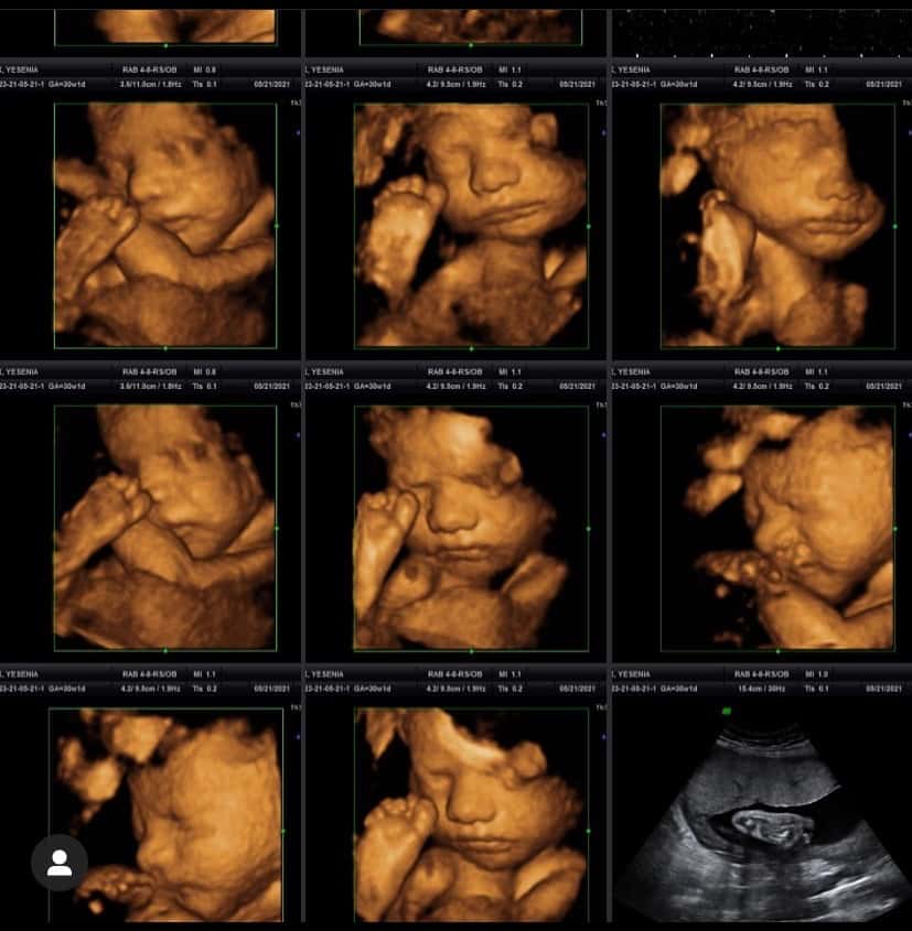
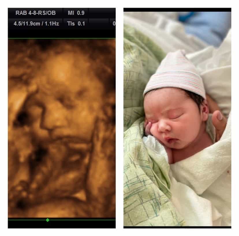
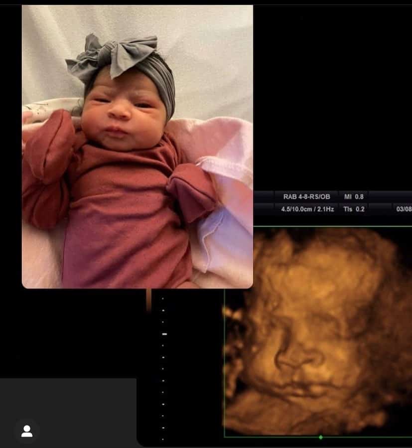
When scrolling through Instagram, we happened to cross paths with the page “all_around_ultrasound” by Cyana Chavez. She is a professional 3D ultrasound fetal photographer and we knew this was worth sharing with our viewers and readers!
But before we get into the baby ultrasound picture gallery, let’s quickly understand the basics of fetal sizes and the developments that a mom-to-be can expect to see in her ultrasounds during her pregnancy weeks.
Baby Ultrasound Pictures in First Trimester - What To Expect?
Here’s what you can expect in your first-trimester ultrasound, week on week, as your baby develops inside the womb. This information is taken and collated from the resources of the American Institute of Ultrasound Medicine (AIUM), Johns Hopkins, and the March of Dimes.
Week 1 - 2
This early in your pregnancy there can be no picture taken to monitor the development of the fetus, however, expect your healthcare provider to count these two weeks to calculate the pregnancy due date, accounting for the first day of your last menstrual period. This is the best reference that your doctor can take to estimate the arrival of the baby.
Week 3
Although in the 3rd week the size of the fetus is not measurable, the baby is getting fertilized. This week the sperm fuses with the egg as it embarks its way from the ovary through the fallopian tube and then into the uterus. Once fertilized, the cells begin to divide rapidly, captured in a sonographer during an ultrasound examination.
Week 4
Week 5
The size of the fetus in week 5 would be 1/18 to 1/16th of an inch, that is the size of the pen dot, while the baby also begins to form the heart and the central nervous system. The ultrasound shows a dark area that is now filled with fluid containing the amniotic fluid.
Week 6
At size 1/6 to 1/4 of an inch, your baby in week 6 takes on a tucked C-shape while forming the head, legs, and umbilical cord with pumping blood through the heart. In the image, you will be able to see big changes this week, your baby will curve inward, umbilical cord in the middle, small buds where the arms and legs will eventually develop.
Week 7
Expect the size of the fetus to be around 1/4 to 1/3 in inches in week 7, that is the size of a pinkie fingernail. This week the baby develops the head, nostrils, and lenses of the eyes. On the other hand, you can expect to see the baby’s development in a bubble within the gestational sac in the ultrasounds, filled with amniotic fluid. This liquid is responsible for giving your baby enough room to grow and develop, while also cushioning your baby-to-be from any external pressure on the abdomen.
Week 8
Size 0.6 inches with the weight, 0.04 ounces, your baby now develops the hands, feet, fingers, but still fused. Also, expect the elbows and ears to start taking shape with little jerky movements that can be seen in the ultrasound in week 8.
Week 9
The size of the fetal in week 9 is almost 3/4 of an inch and weighs 2 grams, except for the development of facial features like eyelids and ears. In an ultrasound, you can expect to see the heart of the baby girl/boy in the blue area, and the red and blue colors within the cord to represent blood going to and from the placenta, picking up oxygen and nutrients from the mama bear!
Week 10
In week 10, the size of the baby is around 1.22 inches with a weight, 0.14 ounces. As per medical research, the baby will develop eyelids, neck, defined fingers, and toes with fully formed ears, although not positioned.
Week 11
At week 11, the fetus is 1.61 inches in length and weighs 0.25 ounces. Expect the little one to develop the chin and neck, defined facial features, and ears higher on the head. In an ultrasound photo, expect to see the tiny fingers and toes forming genitalia, although not visible in images.
Week 12
In week 12, the size of the fetus is 2.13 inches, and its weight 0.49 ounces. The baby will, and continue developing the genitalia, although it isn’t visible on ultrasound.
Week 13
Size almost 3 inches and weight of almost 1 ounce, the baby at week 13 has a functioning kidney and urinary tract with formed fingerprints and developing tooth buds.
Now that we know about the changes and expectations, let’s learn about the ultrasound signs for a baby girl and a baby boy!
Ultrasound Signs!
1) Signs That It's A Girl
Here are the characteristic signs that you can look for to determine if the baby is a girl in an ultrasound:
- The resemblance of a hamburger: The moniker that’s given to the appearance of the clitoris and labia in the ultrasound. Here, the lips of the labia would look similar to that of a hamburger bun, on the other hand, the clitoris, the hamburger patty.
- Sign of sagittal: Each sex has a sagittal sign that can be looked at in the profile view of the fetus, also known as the midline sagittal plane. Also, there is a nub or caudal notch at the end of the spine, pointing downwards at a 10-degree angle.
2) Signs That It's A Boy
Here are the characteristic signs that you can look for to determine if the baby is a boy in an ultrasound:
- The sagittal sign: If the caudal notch points upwards and at more than a 30-degree angle, then be assured that it’s a boy.
- The flow of urine: At times, the flow of the urine could be spotted in a fetus, and in case the flow is moving upwards, then the fetus is a boy!
- Male genitalia: Often visible by weeks 18 to 20, including testicles, scrotum, and penis.


